A stroke is a severe and life-threatening medical condition that occurs when a blood clot cuts off the brain’s blood supply. These clots take time to form and are often detected when it is too late. When a person undergoes this medical emergency, they need to be treated immediately to avoid further complications.
For many years, tissue plasminogen activators (tPA)—drugs that supposedly dissolve the clot—have been the usual treatment for such conditions. This treatment may work for acute strokes, but it is not as effective as it should be for people dealing with a large blood clot.
The longer the time between a stroke and treatment, the worse the patient’s condition could get. Ideally, clot removal must be done within 12 hours of encountering the stroke, or else there might be a smaller chance of survival. Doctors might also find it more difficult to remove the blood clot successfully. Furthermore, the bigger the blood clot is, the more critical the patient’s condition is.
For patients with large blood clots, mechanical thrombectomy (MT) is still the recommended treatment.
What Is a Mechanical Thrombectomy?
A thrombectomy is a surgical procedure of removing blood clots from a person’s artery or vein. The mechanical thrombectomy, on the other hand, is a minimally-invasive procedure wherein a radiologist uses specialized equipment to remove a large clot from the human body. In the form of tiny wire cages, an instrument is inserted into the person’s arteries to extract all the clots at once. A machine called a fluoroscope, which looks like a continuous x-ray, helps make this possible.
For many years, this practice has been seen as useful for patients who experienced minimal brain tissue damage. Still, it was not recommended for patients with severe large ischemic core stroke cases due to lack of clinical evidence.
How the Mechanical Thrombectomy Can Benefit Patients With Large Core Stroke
A study presented by the American Stroke Association in 2019 helped change this narrative. Their presentation suggested that conducting a mechanical thrombectomy specifically for large core stroke patients may provide positive outcomes.
They presented a study that looked into 2,453 patients that underwent clot removal using the said method. 221 of these patients had large core strokes, and 35% of them immediately received functional independence in just three months after the procedure.
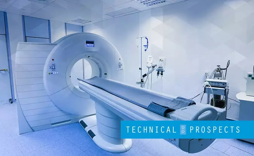
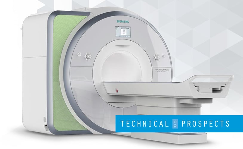
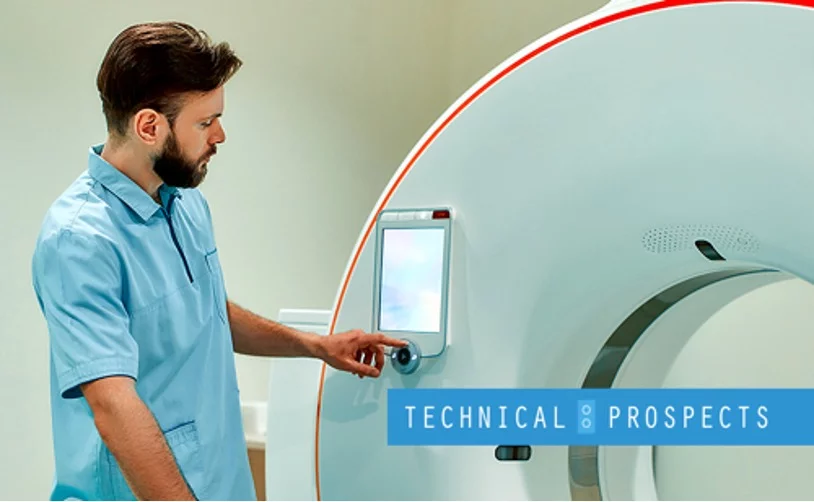
The whole treatment was conducted using CT (computed tomography) imaging and perfusion imaging. CT imaging helped evaluate the flow of contrast dye through the brain’s blood vessels.
The latter imaging is a non-invasive imaging test that shows a person’s blood flow, making the entire treatment more effective. Going through these processes also helped the experts see the extent of the patients’ condition. They were able to identify who among them would be the best candidates for a mechanical thrombectomy.
Conclusion
Thanks to the power of technology and CT imaging machines, saving lives has been made easier. With the help of a perfusion machine, thrombectomy candidates are more comfortable to identify. Conducting the surgical procedure will also be more attainable. With a little more study and more trials, the mechanical thrombectomy’s effectiveness for effectively treating stroke patients might just be proven.
At DirectMed Parts, we believe in the capability of modern machines, especially in science and medicine. We are one of the largest equipment suppliers of MRI and CT parts, and we have the most extensive coil inventory for your MRI and CT needs. If you need a CT or MRI part replacement, we can help. Contact us today!