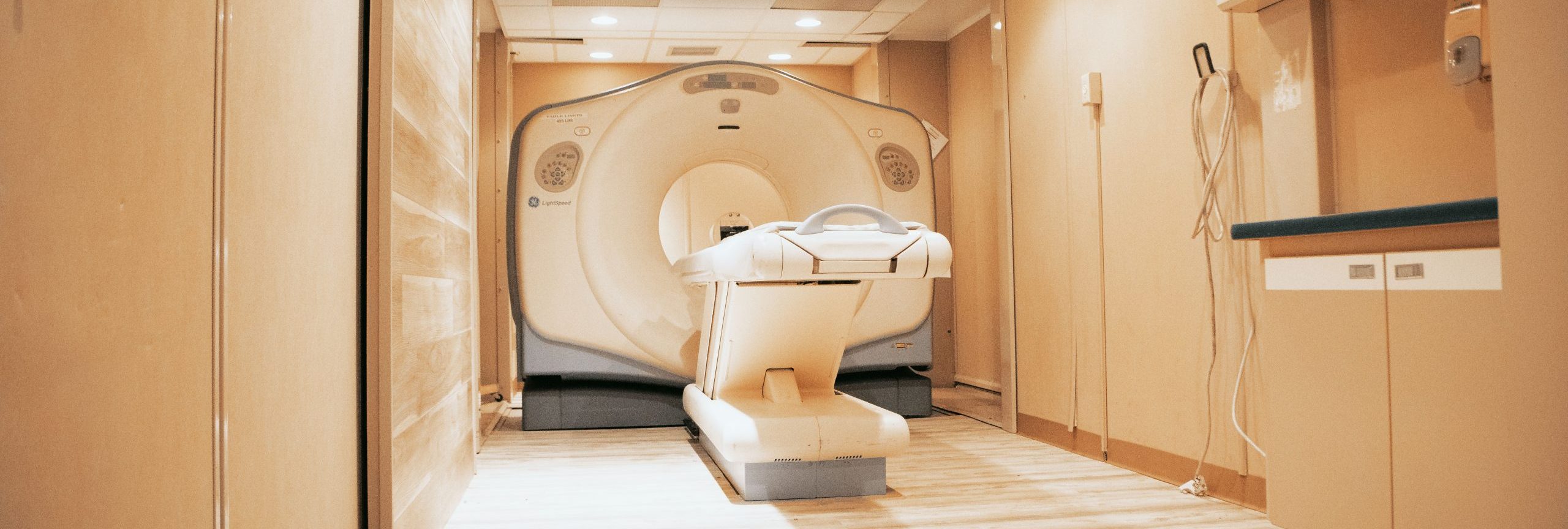
Magnetic resonance imaging systems provide high-quality diagnostic images of patients’ soft tissue and internal organs thanks to their sophisticated technology. It combines high-powered magnets and radio waves to generate incredibly detailed photos, allowing doctors to make more thorough diagnoses and assessments of various conditions.
The clarity and quality of the captured images rely on many aspects, such as the type of MRI system and strength of the produced magnetic field. With a range of low-field to ultra-high field MRI systems on the market, medical professionals can leverage this technology for diagnostic purposes of all kinds. These images’ quality also depends on the imaging techniques used, of which there are two types: T1 and T2.
Understanding How MRIs Work
Knowing the difference between the T1 and T2 imaging techniques requires a basic understanding of MRI functions. These systems use high-powered magnets to produce strong magnetic fields that align the patient’s water molecules’ protons.
These protons absorb the radiofrequency current produced by the MRI system, which also generates a magnetic field. The protons change their rotation after absorbing this energy. When the field is turned off, the protons gradually revert to their original trajectory, an activity known as precession. In the process, they emit a radio signal that MRI parts like the receivers measure, which they use to produce an image of the body part it is scanning.
What is the Difference Between T1 and T2 Techniques?
T1 and T2 refer to the relaxation times used when scanning tissue as an interval between pulse sequences. The T1 technique regulates the rate that protons revert to their regular rotation, and T2 decides the rate at which protons achieve equilibrium or operate at different times. Creating T1 weighted images requires a short time to echo (TE) and repetition times (TR). These refer to the duration between the radiofrequency pulse’s delivery and the capture of the echo signal, and the measured time between pulse sequences applied to an area, respectively. On the other hand, T2 weighted images use longer TE and TR times.
The body is made of different components, so diagnosing a specific condition will require particular setups. It is also tricky to scrutinize an area when other elements of the body are blocking the view. However, combining different MRI sequences with either the T1 or T2 technique will create more detailed imaging.
T1 Weighted Images
T1 weighted images highlight specific elements, making them appear darker or brighter on the scan. Features that appear darker include increased water, like edemas, hemorrhages, tumors, and inflammations; fast-flowing blood; muscles, tendons, and ligaments; bone, cartilage, and calcium; fluid; and abdominal organs.
Meanwhile, elements that look brighter on a T1 weighted image include blood, protein-rich fluids, fat, melanin, subacute hemorrhages. Other factors include high-protein tissue like abscesses, paramagnetic substances, and laminar necrosis of cerebral infarction.
T1 imaging is preferable for creating a picture of the musculoskeletal system and brain structure since the bone marrow contains a lot of fat, which appears bright on the scan. This feature makes it useful for diagnosing conditions like multiple sclerosis and leukemia.
T2 Weighted Images
T2 imaging is similar to the T1 technique because certain elements appear brighter or darker on the scan. Elements that appear dark on a T2 weighted image include calcium, the liver, air, tendons and ligaments, rapidly flowing blood, adrenals, and cartilage.
Meanwhile, elements that look brighter on a T2 weighted image include fluid, kidneys, the pancreas, muscles, the bladder, bile, and the gallbladder. T2 weighted images often highlight water and fluids, making it excellent at identifying areas of inflammation. It’s also popularly used in pathology, such as assessing the kidneys’ or looking for signs of disease.
Conclusion
The T1 and T2 imaging techniques are available when performing an MRI scan, allowing you to choose the method that highlights the elements you are looking for when assessing a patient. Understanding how each technique works and the features they highlight enables you to make the best choice between T1 and T2 imaging, providing an accurate and high-quality scan of the problem area.
DirectMed Parts is the most reliable and trustworthy source for medical imaging parts and services. We specialize in CT and MRI parts and coils while offering MRI coil repair. Contact us today to learn more about what we can do for your medical imaging system!