A person showing signs of having a stroke must undergo brain imaging within 25 minutes of emergency room admission. Delays may reduce effectiveness in administering the proper treatment.
When it comes to a stroke, speed and time are significant factors for ensuring the patient’s survival and minimizing the extent of brain damage. Computed tomography (CT) scan is the standard test to assess a stroke. While this procedure is quick and easily accessible, the American Academy of Neurology revealed that magnetic resonance imaging (MRI) scans are better at detecting stroke damage than CT scans. To learn more about MRI and its importance in treating a stroke, use this article as your guide.
How Does a Stroke Occur?
Stroke is the third leading cause of death and the leading cause of serious, long-term disability, causing more than 140,000 people to die yearly. A stroke happens when blood flow to a part of the brain gets blocked by a clot or broken blood vessel. When this occurs, cells in the brain have no means to receive the constant supply of oxygenated blood they need, causing permanent brain damage.
What Are the Different Types of Stroke?
Stroke has two different types, and each requires a unique treatment. A hemorrhagic stroke occurs when a blood vessel in the brain ruptures and causes blood to leak into the brain, while an ischemic stroke is the most common type of stroke that happens when a blood vessel gets blocked by a blood clot or arteries.
Why Is MRI Used for Stroke?
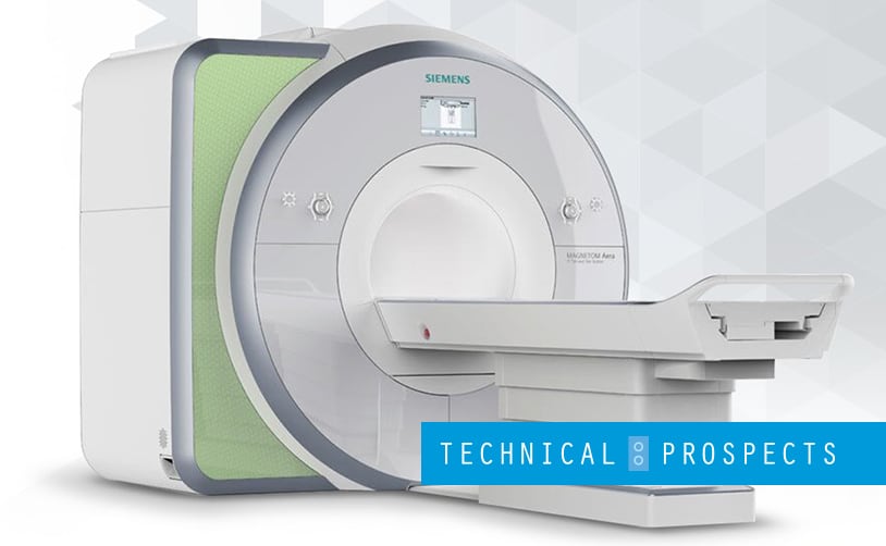
An MRI is a medical imaging test that uses radio waves and a magnet to produce a highly detailed image of the brain. This machine can detect various brain and blood vessel abnormalities.
Compared to X-rays and CT scanners, it can visualize even the smallest differences between tissues or those located in regions of the brain that cannot be detected by CT. It can also distinguish ischemic lesions and identify pathologies that resemble a stroke. In addition, it can provide information about the size and location of the stroke event.
MRI offers various benefits, including the absence of radiation, whole-brain coverage, and a safe contrast applied in lower doses. Its technique, particularly the arterial spin labeling (ASL), allows for basic perfusion imaging without administering contrast agents.
How Is MRI Used to Detect a Stroke?
When assessing a stroke patient using MRI, it’s important to determine whether the patient has experienced a hemorrhagic or ischemic stroke. An MRI of the head is generally the first test conducted.
MRI images of the human brain are usually taken in three orientation displays: coronal, sagittal, and axial. These orientations show a slice dividing the head into the front and back halves, left and right halves, and upper and lower halves. They provide radiologists with a detailed 3D view of the body part.
Conclusion
MRI is a vital tool for detecting, diagnosing, and treating a stroke. It is an important machine for healthcare facilities. At present, MRI is expected to be continuously effective in monitoring stroke patients. This is especially true as technology advances when it can have stronger magnets and faster scan times and be used in emergency settings.
Since speed is essential in saving the lives of stroke patients, remember the information above, invest in a quality MRI scanner, and seek repair and maintenance when needed.
If you need MRI parts, coil repair, and other related services, you can count on us at DirectMed Parts. We are your reliable source for medical services and imaging parts. Contact us to maintain the excellent condition of your machine and provide your patients with better healthcare!