Magnetic resonance imaging is a powerful tool used in the medical field to produce highly-detailed, high-quality, and high-resolution images of a person’s brain. Advanced MRI parts work to capture more comprehensive details on the brain’s anatomical structure, all without the dangers of radiation. MRIs are suitable for imaging soft tissues in the body, including the brain for better diagnosis and research. Understanding just how it can benefit neurology can lead to future breakthroughs in medical treatments as technology advances. This article discusses the ways that the specialization of neurology can
Types of MRI in Neurology
There are different types of MRIs that are valuable for studying and diagnosing the brain. First, there is the functional MRI. This observes the brain structures and scans the organ while patients are asked to perform cognitive tasks, which can include puzzles, mathematical solutions, or responding to other stimuli such as sounds or lights. The fMRI allows doctors and patients alike to review the visualization of neural activity in the brain and spinal cord, making it especially useful for neuroscientific research.
Diffusion tensor imaging, on the other hand, is quite unique with its imaging modalities. This can visualize the more detailed tracts in the brain, making it an excellent tool to identify damage and disconnection. Conventional MRIs will not be able to detect conditions such as traumatic brain injury, but a DTI can.
MRI Neuroimaging
When using MRI imaging, there’s a whole list of opportunities that opens up for patients and doctors. MRI can penetrate soft tissue, making the data and images it gathers incredibly valuable to the patient’s diagnosis and treatment. Examples of patients who can benefit greatly from this technology include those with multiple sclerosis (MS), stroke, brain tumors, and other abnormalities or brain diseases.
Bear in mind that an MRI will not isolate the specific location of the brain that controls a certain function. Fortunately, doctors are given enough information to map out the functional areas, especially if there’s a need for surgery and other treatments.
Magnetic Resonance Angiography (MRA)
There are different types of procedures related to the MRI that can spot and diagnose specific types of neurological conditions. One such example is Magnetic Resonance Angiography (MRA). This technique can be used to evaluate the blood flow in arteries and other chemical imbalances in the body. It can address chemical abnormalities in body tissues such as HIV infection of the brain, tumors, Alzheimer’s disease, head injuries, and more.
MRI Improves Visualization
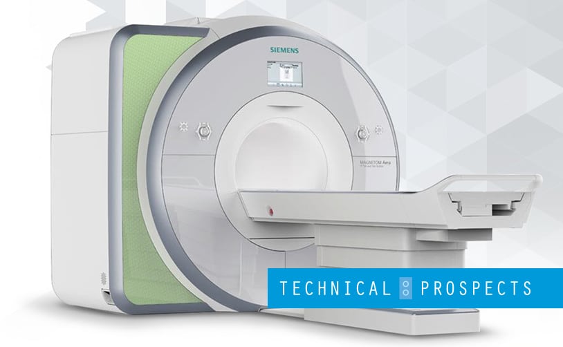
MRI technology has come a long way since it was introduced into the medical world. It has continued to evolve and advance to produce sharper and clearer images in less time. Some of the most powerful MRI machines use 7T level magnetic fields that can visualize the brain in impressive detail using enhanced contrast mechanisms. One such mechanism is the oxygen-level dependent and flow-dependent contrasts. Neurologists can study 7T MRI scans to see visible lesions and brain abnormalities much more clearly. For instance, MRI scanners of this strength can identify which specific part of the brain epileptic seizures come from. It can also visualize brain tumor pathologies and neuron loss, among other things.
Apart from visualization, MRI technology has also improved in terms of brain scan speed, accuracy, and ease. Small and subtle lesions can be examined in great detail using 3D volume scans with thin-slice, SNR-rich studies. 3D arterial spin labeling also provides opportunities to rule out focal or global perfusion defects or other cerebrovascular diseases. MRIs also provide automated brain exams, which reduces human error and inconsistency and reduces the workload.
Artificial Intelligence and Neurology
AI has started to play a greater role in neuroimaging during the last few years. Any errors found through interpretive and perceptual errors are greatly reduced with computer analysis of highly augmented images.
Conclusion
As time goes on, the future for MRI imaging capabilities in specific medical fields such as neurology looks bright. More advances will push for stronger MRI magnetic fields and color contrast methods. This is one way to result in accurate diagnosis for neurological disorders, better treatment, and a much deeper understanding of the complex brain and how diseases develop. Whether your team needs to change out the mechanical MRI coil for repair or decide to replace your existing MRI unit completely for an upgrade, knowing where to get quality parts and services is a must.
At Direct Med Parts & Service, we are the most knowledgeable and trusted source for parts relating to medical imaging and services. We prioritize the delivery of our CT and MRI parts and coils to service professionals promptly to ensure efficient medical services. Get in touch with us today for the MRI and CT services you require!
